AI Diagnostics in Women's Reproductive Health
AI is reshaping how doctors diagnose endometriosis and PCOS, but validation gaps remain.
Here is the number that reframes everything: Silicon Valley Bank tracked USD 2.6 billion deployed into women's health in 2024. That is a record, and it arrives in a sector that, for most of its history, could not attract institutional capital at anything approaching that scale. Think of it as a field that spent decades knocking on doors only to find the whole street finally opening up at once.
The private money is not moving alone. The White House committed $500 million in 2024 to women's health research through NIH and ARPA-H. Dollar figures matter less than what federal research infrastructure does to a clinical domain when it formally adopts one: it changes what gets studied, what journals publish, and how regulatory reviewers prioritize incoming applications. That downstream legitimization is often worth more than the grant dollars themselves.
The FemTech market, valued at USD 8.56 billion in 2025 and forecast to reach USD 18.98 billion by 2031 at a 14.20% compound annual growth rate according to Mordor Intelligence, is growing fast enough that Flo Health's USD 200 million fundraise and unicorn status in 2024 barely registered as surprising. Consumer women's health technology has crossed into commercial viability.
What the funding figures cannot tell you is the distinction between consumer-facing cycle apps and clinical diagnostic tools. Period tracking serves real needs. It is not, however, AI detecting endometriosis from ultrasound or stratifying preeclampsia risk in the first trimester. The clinical diagnostic layer is where the evidence base is actively developing, and where the funding is most consequential for patient outcomes. The money and the research have yet to converge, and that gap is where most of the interesting work is happening.
Research volume is its own momentum signal. A 2025 review found 1,768 published articles on AI in women's health across PubMed and MEDLINE for the five-year period ending May 2024, with 207 meeting criteria for substantive review. That compression from early exploration to active clinical investigation is not gradual. It is abrupt, and we are in the middle of it.
How AI Diagnostic Tools Actually Work in Reproductive Medicine
The tools are not one thing, and conflating them creates real confusion about what is actually possible.
Two broad families of method exist, and the distinction determines which problems each family can solve. Traditional machine learning, covering support vector machines, random forests, and logistic regression, works well when input data is structured: blood markers, clinical history, lab results, demographic variables. These models learn which feature combinations best predict an outcome and train on relatively modest datasets. Deep learning, particularly convolutional neural networks, handles unstructured data: images, video, waveform patterns. When your input is a colposcopy image or a time-lapse of a developing embryo, deep learning detects features a human reader does not consciously register and would consistently miss across thousands of cases even if she tried. In other words, traditional machine learning is like a detective who reads the case file; deep learning is like one who watches the surveillance footage frame by frame.
Input data types vary considerably across reproductive medicine applications. Imaging, ultrasound, colposcopy, time-lapse embryo video, is the substrate for some of the most clinically advanced tools. Other applications draw on biochemical markers, ECG signals, electronic health records, and genetic and proteomic data. Multimodal approaches, the ones combining several of these streams, consistently produce the most robust predictions. No single data type fully characterizes a complex condition.
At its best, what AI contributes is pattern recognition across high-dimensional data at a scale and consistency no individual clinician can sustain. A specialist brings deep expertise but also fatigue, variability across readings, and the cognitive bandwidth limits that come with being human. A well-trained model applies the same decision logic to every case it sees. That consistency is underrated.
Four metrics recur throughout the evidence base. Accuracy is the proportion of cases classified correctly. AUC, area under the curve, measures how well a model distinguishes positive from negative cases across all possible decision thresholds; a score of 1.0 is perfect, 0.5 is chance. Sensitivity is the model's ability to correctly identify true positives. Specificity is its ability to correctly identify true negatives. High sensitivity with lower specificity means fewer missed diagnoses and more false alarms. Balancing them is a clinical and regulatory judgment, not a purely technical one, and the right balance differs by condition.
One limitation applies across nearly every tool currently in development: most validation is retrospective and single-center. Models trained and tested on data from one institution, one scanner type, one patient population frequently perform measurably worse when deployed somewhere new. Every strong accuracy figure in this field carries that caveat, and the research community knows it. Whether they communicate it clearly is another matter.
Endometriosis: Shortening a Diagnostic Delay That Averages Years
The diagnostic problem with endometriosis is structural, and it has been structural for a long time. The condition has a complex, incompletely understood etiology. Its symptoms overlap with other pelvic disorders. There is no reliable non-invasive test. The gold standard remains laparoscopic surgery: an operative procedure requiring anesthesia, a surgeon, and recovery time. The result is a diagnostic odyssey that spans an average of several years, years filled with pain, treatment attempts aimed at the wrong targets, and progressive tissue damage accumulating the entire time. It is, in the bleakest sense, a condition that hides in plain sight.
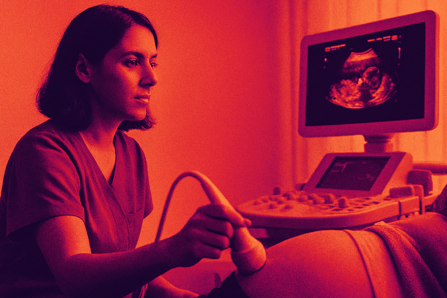
AI applied to ultrasound is the most developed non-invasive alternative. Deep learning models evaluated in reviewed studies achieved accuracy in the range of 0.89 to 0.93, AUC around 0.90, sensitivity between 0.78 and 0.92, and specificity between 0.74 and 0.89, according to a 2025 analysis in Contemporary OB/GYN. Near-expert-level results from a non-invasive scan. In practical terms, that means AI-assisted ultrasound can confirm or rule out endometriosis for a meaningful proportion of patients before anyone picks up a laparoscope.
A clinical trial completed at University Hospital Tübingen in June 2025 tested AI-based real-time detection of endometriosis lesions from endoscopic image and video data during surgery. The intraoperative framing matters. This is not AI replacing the surgeon; it is AI operating alongside the surgeon, flagging lesions that might otherwise be missed in the moment. That is a distinct use case from pre-operative screening, and both are genuinely useful.
The access argument follows directly. Expert sonographers who can reliably identify endometriosis on ultrasound are concentrated in specialist centers. An AI system performing at expert level reduces that operator dependence, which extends the tool's clinical reach to rural and under-resourced settings where those specialists simply do not exist. Extending interpretive capacity to where it is most absent is as consequential as any accuracy metric.
What remains unresolved is generalizability. Most studies are small and retrospective. Performance across different ultrasound equipment manufacturers, different imaging protocols, and diverse patient populations has not been rigorously established. Those are the validation studies the field needs before any of this becomes standard of care, and they are not yet complete.
PCOS: Applying AI to a Condition Whose Diagnosis Is Still Contested
Polycystic ovary syndrome affects somewhere between 4% and 20% of reproductive-age women globally. That range is not a rounding problem; it tells you how inconsistently the condition is identified. More than 66 million women are affected worldwide. In the United States, PCOS-related symptom management cost nearly $8 billion in 2020, according to data published in Frontiers in Endocrinology. Despite that scale, more than one-third of women with PCOS experience a diagnostic delay exceeding two years, and that figure is almost certainly an undercount.
AI and machine learning have been applied to PCOS across clinical examination data, electronic health records, and genetic and proteomic inputs. A 2023 systematic review that screened 135 studies and included 31 found AI interventions drawing on all of these modalities. A 2025 review from NIH and NIEHS confirmed that AI and ML programs can successfully detect and classify PCOS across data types.
Here is the complication that does not get enough attention. PCOS is a diagnosis of exclusion, established by criteria that clinicians apply inconsistently in practice. When an AI model trains on labeled data, it inherits whatever ambiguity existed in how those cases were labeled. A model trained on inconsistently diagnosed PCOS cases will reproduce that inconsistency at scale, with the added problem that it will do so confidently and uniformly. You can say the model has no knowledge problem — it has an inheritance problem. This is not a reason to dismiss AI's role. It is a reason to be precise about what problem the AI is actually solving: it can identify consistent patterns within a heterogeneous condition better than any individual clinician, but it cannot adjudicate the upstream definitional disagreement the field itself has not resolved. The model reflects its training labels. No more, no less.
Solence's July 2025 launch of an AI-powered digital therapeutic specifically for PCOS signals movement beyond detection into ongoing management, treating the condition as chronic and requiring longitudinal support rather than a single diagnostic event. That reframing is more consequential than any individual accuracy metric, because it asks a harder and more useful question: what does it mean to actually manage this condition over time?
Preeclampsia Prediction: Catching a Dangerous Condition in the First Trimester
More than 76,000 women die every year from preeclampsia and related hypertensive disorders of pregnancy. Let that number sit for a moment. Traditional screening tools miss up to 66% of patients who go on to develop the condition, according to a 2025 analysis published in PLoS ONE. That failure rate is not a gap in the system. It is the system, and it is what makes first-trimester AI-based prediction worth taking seriously.
The intervention window is early pregnancy. Preeclampsia manifests in the second half, but the biological processes causing it begin in the first trimester, when effective preventive intervention is still possible. A 2025 AI predictive framework integrates maternal demographics, biophysical parameters, and first-trimester biochemical markers into a composite risk score targeting precisely that early window. No single first-trimester marker is sufficiently predictive on its own; the biology requires combinations of markers processed by a model trained on large datasets. The multimodal approach is not a design preference. It is what the underlying physiology demands.
An emerging modality deserves attention even at an early stage: AI models that detect preeclampsia risk from ECG signals in point-of-care settings. ECGs are inexpensive, non-invasive, and available in health settings where specialist obstetric screening is absent. If ECG-based risk stratification validates in larger studies, the access implications extend well beyond what specialist-dependent screening can ever reach.
A 2025 PRISMA systematic review evaluated machine learning models across 11 studies comprising 116,253 pregnancies. The field is still accumulating the prospective evidence needed for broad clinical deployment. An active trial at Istituto Clinico Humanitas is examining a multiomic AI approach investigating the maternal microbiota's role in immune response and metabolism in hypertensive disorders. That direction signals movement toward biological mechanism rather than pattern matching on clinical variables alone, which matters considerably for long-term generalizability. Pattern recognition on surface variables can fail quietly when the underlying biology shifts; models grounded in mechanism tend to be more durable.
IVF Embryo Selection: Where AI's Promise and Its Limits Are Both Visible
More than 100,000 babies were born in the United States through IVF in 2024. Success rates drop to around 10% or lower for people over 40, according to Scientific American. That outcome gap is what AI-assisted embryo selection is trying to close, and for anyone who has been through IVF, it is not an abstract problem.
The application works like this: deep learning and computer vision analyze time-lapse video of embryos developing in culture. Platforms including ERICA, iDAScore, and IVY score embryos based on morphokinetic features, replacing the subjective visual grading embryologists have traditionally used. A systematic review of 27 studies found an average AUC of 0.91 for treatment outcome prediction, with 17 of those studies using deep learning. In controlled settings, those are strong numbers.
The cautionary data point deserves equal weight. A 2024 randomized controlled trial conducted in the UK and Hong Kong with nearly 1,600 participants found live birth rates of 33.7% with AI-assisted time-lapse imaging versus 33% without. Preliminary data from the Matris AI system, drawn from 150 cases, showed up to 15% improvement in implantation rate over traditional methods, but the investigators explicitly noted the need for validation in larger, multicenter trials.
The pattern across this field is consistent: AI performs well in retrospective studies and small prospective studies; large randomized controlled trials have not confirmed proportionate gains in outcomes patients actually care about. Strong AUC scores and live birth rates are measuring different things. One measures a model's ability to rank embryos. The other measures whether a baby comes home. Commercially deployed tools are not always forthcoming about which number they are citing, and patients navigating high-stakes, expensive treatment cycles deserve to know the difference.
Cervical Cancer Screening: The Application Where AI Has Reached Clinical Deployment at Scale
Gynecologic cancers affect more than 1.2 million women globally each year. Cervical cancer is the most preventable of them, provided screening actually reaches the women who need it. In much of the world, it does not, because the workflow depends on trained cytopathologists who are not available at the required scale.
AI-enhanced Pap smear analysis and colposcopy have achieved up to 95% accuracy in cervical cancer screening, based on analysis of 75 peer-reviewed articles published between 2017 and 2024. In February 2024, Hologic launched the first FDA-approved digital cytology system: the Genius Digital Diagnostics System with the Genius Cervical AI algorithm. FDA clearance for a diagnostic AI tool in oncological screening demands a level of evidentiary rigor that laboratory performance numbers alone do not satisfy. Regulatory approval is a meaningful signal here, not just a commercial one.
The deployment that makes this case genuinely distinctive happened in China's Hubei province, where an AI-plus-cloud cervical screening program covered 1,704,461 individuals between July 2022 and January 2023. Nothing else in this space comes close to that real-world validation. There is a considerable difference between a tool that performs well in a study and one that has been stress-tested at population scale, and this one has been.
Cervical cancer is AI's most tractable diagnostic target because of a specific alignment between what the technology does well and how the clinical workflow is structured. The screening task is image in, binary classification out — as clean a problem as a lock waiting for the right key. Ground truth is well-defined by biopsy. The imaging modality is standardized. The cost of a missed positive is catastrophic. These features make the application ideal for computer vision in a way that more heterogeneous diagnostic problems are not. The tool substitutes for specialist cytopathologists in settings where they are absent, which is exactly where screening coverage gaps are most severe. That alignment between technical capability and clinical need is rarer than it sounds, and it explains why this application is years ahead of the others.
What the Shift Toward AI Diagnostics Means for Patients and Clinicians in Practice
What connects endometriosis ultrasound, first-trimester preeclampsia prediction, embryo scoring, and cervical cytology is not that AI is replacing clinical judgment. It is that AI is reducing the operator dependence and specialist bottleneck that currently determine who receives accurate diagnosis and who does not. Expert-level interpretation of a pelvic ultrasound, an endoscopic video, or a cytology slide is unevenly distributed across geography, institution, and economic circumstance. These tools can extend that interpretive capacity to settings where it would otherwise not reach, and that extension is the actual value proposition.
For patients, earlier detection means less invasive treatment, less time living with symptoms that have no name, and, in obstetric cases, lower risk of outcomes that kill. For clinicians, the appropriate framing is AI as a second reader or risk-scoring layer, not an autonomous decision-maker. Regulatory approval, clinical adoption, and liability questions the field has not yet resolved all push toward that framing, even when the technology's performance technically supports something more autonomous.
Low-resource and rural settings stand to gain disproportionately if AI can deliver expert-level diagnostic interpretation beyond specialist centers. That benefit only materializes if deployment decisions follow clinical access needs rather than commercial opportunity. There is no structural guarantee those two will align, and the history of medical technology suggests they often do not, at least not at first. Early adopters in well-resourced settings tend to benefit first, even when the population most likely to benefit from the technology sits somewhere else entirely.
Regulatory pathways for multimodal AI diagnostic tools are still developing. Training data skewed toward well-represented populations can embed diagnostic bias that only becomes visible when the model encounters a different demographic context. Liability frameworks for AI-assisted misdiagnosis are genuinely unresolved, and that unresolved status creates institutional caution among clinicians who would otherwise move quickly.
Most applications in this space have strong retrospective evidence and incomplete prospective validation. Cervical cancer screening has crossed into large-scale real-world deployment. The others are at various stages, accumulating the multicenter prospective trial data that will determine whether early research results hold at population scale. The trajectory is real. Most of the proof is still coming.